Research

From worms to human cells

Every animal must tell friend from foe among the microbes it meets. We study how cells sense infection, which genes they switch on, and how the lysosome and the transcription factor TFEB tie these steps together.

Our approach

Three systems, one set of questions

We move between a simple animal and human cells. Worms let us search the whole genome quickly; immune and gut cells let us test whether what we learn holds in mammals.

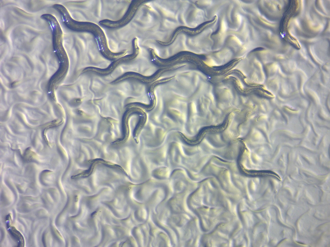
Many small roundworms crawling on the surface of a culture plate

C. elegans

A transparent roundworm about one millimeter long. It eats bacteria, so its gut meets microbes every day, and its genes can be switched off one at a time.

Colorized electron micrograph of a green white blood cell engulfing round purple bacteria

Macrophages

Immune cells that engulf and destroy bacteria. We study mouse macrophages and the human THP-1 macrophage cell line.

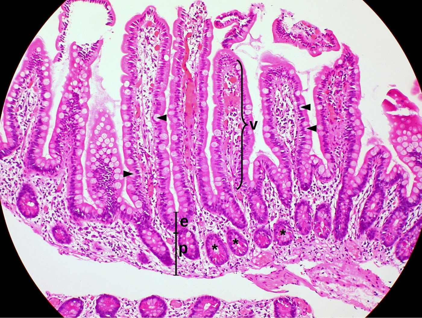
Pink and purple stained section of the small intestine showing finger-like villi

Intestinal epithelial cells

The single layer of cells that lines the gut and keeps microbes out. We study them in mice and in human cells.

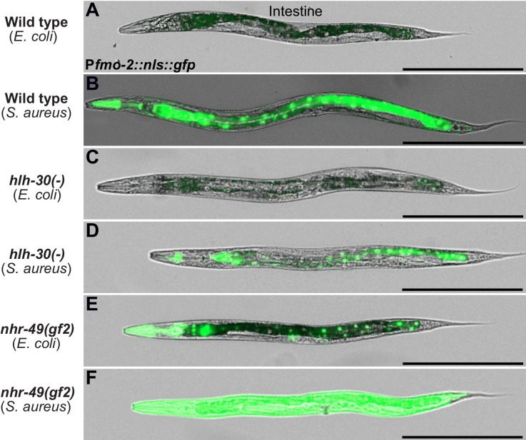
Six microscope images of whole worms; in infected wild-type worms the gut glows bright green
An infection-induced defense gene (fmo-2, green) switches on in the worm gut. From Wani et al., eLife 2021, Figure 4, figure supplement 1, CC BY 4.0.

Theme 1

TFEB, a master switch for defense

When an animal is infected, its cells must turn on the right defense genes at the right time. We found that TFEB, called HLH-30 in worms, is one of the main switches.

What we found

  • In worms infected with Staphylococcus aureus, HLH-30 switched on soon after infection and drove close to 80% of the animal's response, including genes that kill microbes and genes for autophagy (cell recycling). TFEB was also needed to turn on several inflammatory genes in mouse macrophages. PMID 24882217
  • Removing HLH-30 erased almost all genes turned on by infection but not by starvation. One of the few left, the defense gene fmo-2, was essential for survival; it needed a second regulator, NHR-49/PPAR-α, to switch on. PMID 33978570
  • HLH-30 also turns on NHR-42, which acts as a brake: worms lacking NHR-42 survived infection better and made more antimicrobial genes. PMID 36860863
  • Our review describes TFEB and its relative TFE3 as key regulators of innate immunity and inflammation. PMID 31959514

Key papers: PMID 24882217, PMID 33978570, PMID 36860863, PMID 31959514
Colorized electron micrograph of a green white blood cell engulfing round purple bacteria
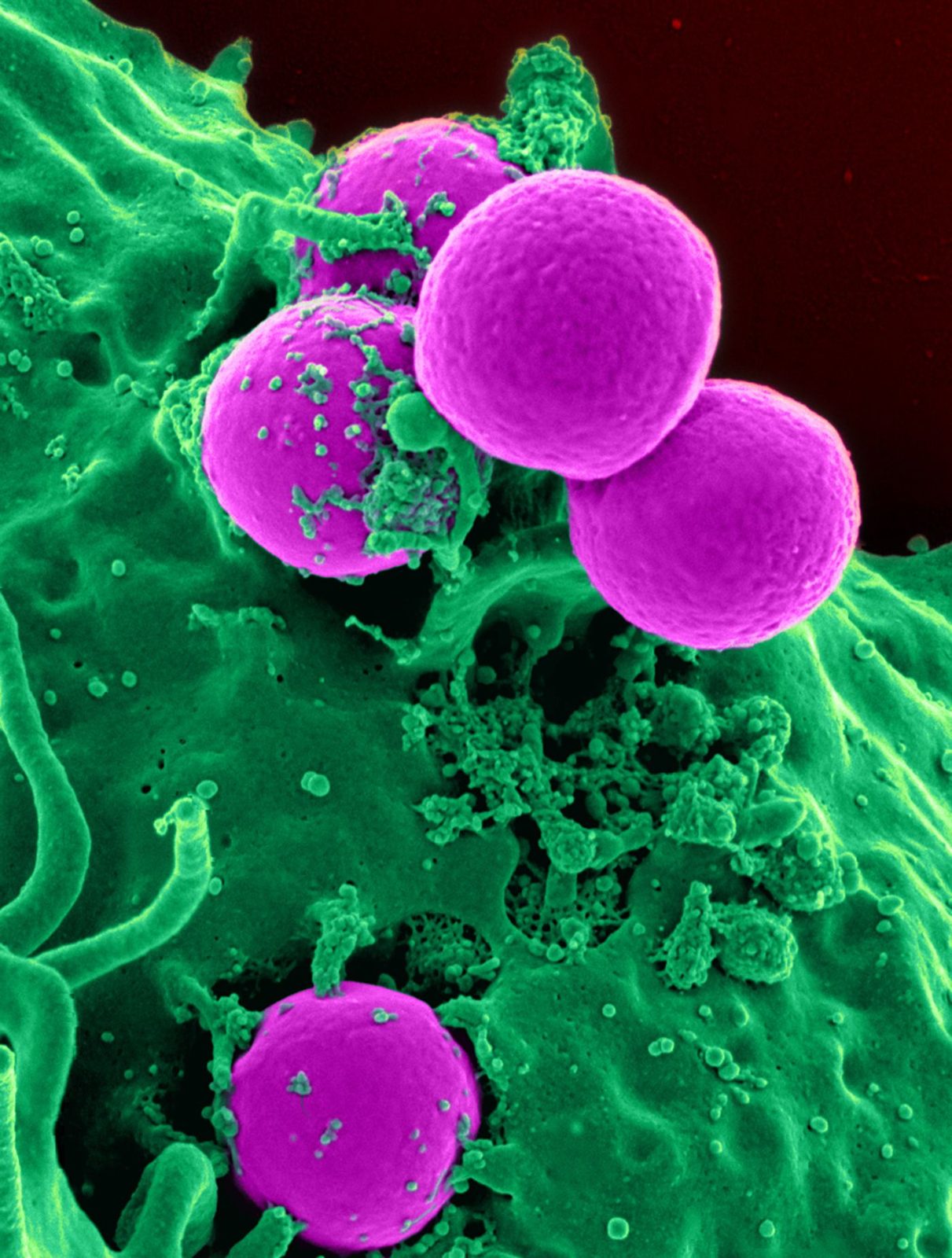
A human white blood cell (neutrophil, green) engulfing MRSA bacteria (purple). Image: NIAID/NIH, Wikimedia Commons, public domain.

Theme 2

What flips the switch in immune cells

Macrophages swallow and destroy bacteria. We trace the chain of signals that links this contact, and nerve-like chemical signals, to TFEB turning on inflammatory genes.

What we found

  • In worms, TFEB activation needed a chain of three signaling proteins that ends with protein kinase D. A similar chain worked in mouse macrophages infected with Salmonella, a pathway kept across a billion years of evolution. PMID 27184844
  • When macrophages swallow bacteria, a burst of reactive oxygen made by the NOX/PHOX enzyme turns on TFEB through CD38, the calcium messenger NAADP, and calcineurin. Macrophages lacking TFEB and TFE3 could not mount an inflammatory response to bacteria. PMID 33818279
  • Turning on the α7 nicotinic acetylcholine receptor moved TFEB into the nucleus, expanded lysosomes, and switched on inflammatory genes. This needed the lysosome calcium channel MCOLN1 and calcineurin. PMID 40435320

Key papers: PMID 27184844, PMID 33818279, PMID 40435320

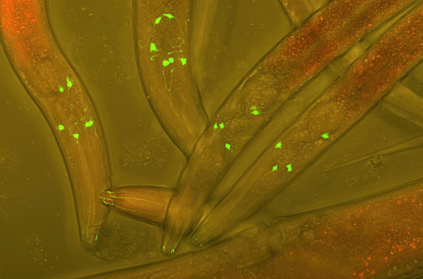
Microscope image of transparent roundworms with glowing green nerve cells
Live C. elegans with fluorescent (GFP) neurons. Image: Heiti Paves, Wikimedia Commons, CC BY-SA 3.0.

Theme 3

How nerves shape defense in the gut

The nervous system and the immune system talk to each other. In worms we can watch that conversation in a living animal.

What we found

  • During infection, neurons release acetylcholine, which acts on the gut lining through muscarinic receptors. This turns on Wnt signals that switch on antimicrobial genes. PMID 29768179
  • With the Srinivasan lab, we found that S. aureus infection damages the sensory branches of the worm's pain-sensing ASH neurons, which we call pathogen-induced neurite pathology (PaIN). Removing HLH-30/TFEB cut how often this happened by about half. PMID 41327171
  • Our review summarizes how the worm nervous system turns gut defense genes on and off. PMID 31790812

Key papers: PMID 29768179, PMID 41327171, PMID 31790812

Colorized electron micrograph of clusters of round yellow bacteria on a red human immune cell
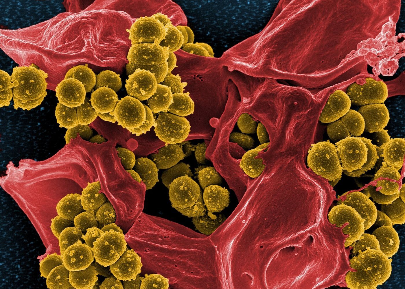
Staphylococcus aureus (MRSA, yellow) on a dead human neutrophil (red). Image: NIAID, Wikimedia Commons, CC BY 2.0.

Theme 4

Friend or foe: gut microbes and infection

The microbes in an animal's gut can help or harm it, and the same microbe can do either, depending on the host's defenses.

What we found

  • Most members of CeMbio, a standard 12-species worm gut community, switched on HLH-30/TFEB and shortened the lives of worms lacking it. Host defense masks this "cryptic virulence." PMID 38623070
  • Worms lacking EGL-9, which restrains the oxygen-sensing factor HIF, were more susceptible to S. aureus but more resistant to Pseudomonas aeruginosa. HIF can tailor the response to each pathogen. PMID 22792069
  • Our review proposes that reactive oxygen made during infection upsets the cell's protein upkeep and kills worms whose infections do not clear. This is a model we are testing. PMID 32898751

Key papers: PMID 38623070, PMID 22792069, PMID 32898751

Pink and purple stained section of the small intestine showing finger-like villi
Lining of the small intestine, stained with hematoxylin and eosin. Image: Ed Uthman, Wikimedia Commons, CC BY 2.0.

Theme 5

TFEB protects the gut lining

The cells that line the intestine form a barrier that must withstand injury and repair itself. We asked whether TFEB helps.

What we found

  • Mice lacking TFEB only in the gut lining looked largely normal but had a defect in the granules of Paneth cells, a specialized gut cell. PMID 29066772
  • These mice made less ApoA1, a lipoprotein that is reduced in people with Crohn's disease, and they developed worse colitis after chemical injury to the gut. PMID 29066772

Key papers: PMID 29066772

Close-up fluorescence image of human cells with many small red dots, the lysosomes, clustered around blue nuclei and green fibers
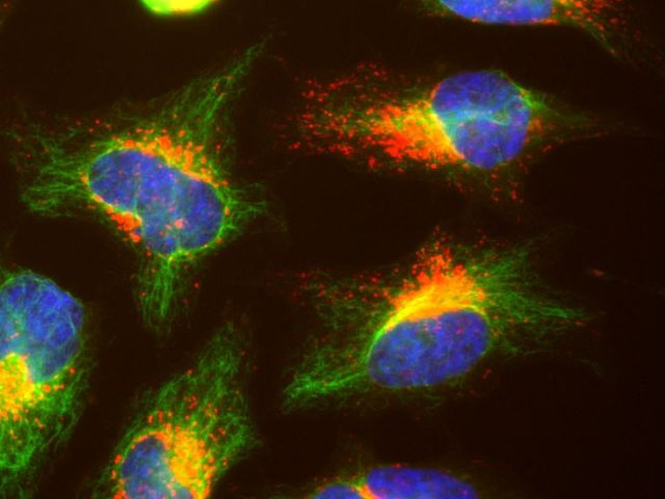
Close-up of human cells: lysosomes stained for LAMP1 (red), vimentin (green), and DNA (blue). Image: GerryShaw, Wikimedia Commons, CC BY-SA 4.0; cropped.

What comes next

Current directions

Our newest work asks how the cell's organelles and its immune defenses work together.

  • Lysosomes and peroxisomes. How do these two organelles change during infection, and how does TFEB/HLH-30 coordinate them?
  • Complement-driven phagocytosis. How do human macrophages eat microbes tagged by complement proteins, and what happens inside the cell next?
  • Stress resilience in the gut lining. How does TFEB help human intestinal epithelial cells withstand stress?

Funding

Our funding

We are grateful to the National Institutes of Health and the National Science Foundation for supporting our work.

Current

  • NIH NIGMS R35 GM149284 (Maximizing Investigators' Research Award). Mechanisms of the gut-brain axis that regulate innate immunity. 2023 to 2028.

Past

  • NIH NIAID R21 AI169842. Flavin-containing monooxygenases as novel innate immunity effectors. 2023 to 2025.
  • NIH NIGMS R01 GM101056. Role of MiT transcription factors in host defense against bacterial infection (2012 to 2016), renewed as Nervous system control of intestinal host defense mediated by TFEB (2017 to 2024).
  • NSF IOS 1457055 and 1656925. Neuronal Wnt signaling and intestinal host defense in nematodes. 2015 to 2018; the award moved with the lab from Massachusetts General Hospital to UMass Chan.
  • NSF IOS 1566013. Support for the Fourth Topical Meeting on C. elegans Pathogenesis, Aging, Stress, Small RNA, and Metabolism. 2016.

Sources: NIH RePORTER and the NSF Award Search. Fellowships are listed with honors on the People page.